
Dr. Raveesh Sunkara

What Is Brain Aneurysm?

Schedule ConsultationCase Review

Reviewed personally by Dr. Sunkara · Written for Brain Aneurysm & AVM · Updated September 2026

Overview

How a Brain Aneurysm Forms

Quick Answer

A brain aneurysm is a weak, bulging spot on a blood vessel wall inside the brain, shaped like a small balloon filled under pressure. Most stay silent for years and are found only on a scan done for something else. A related but separate condition, an AVM, is a tangle of blood vessels rather than a single bulge.
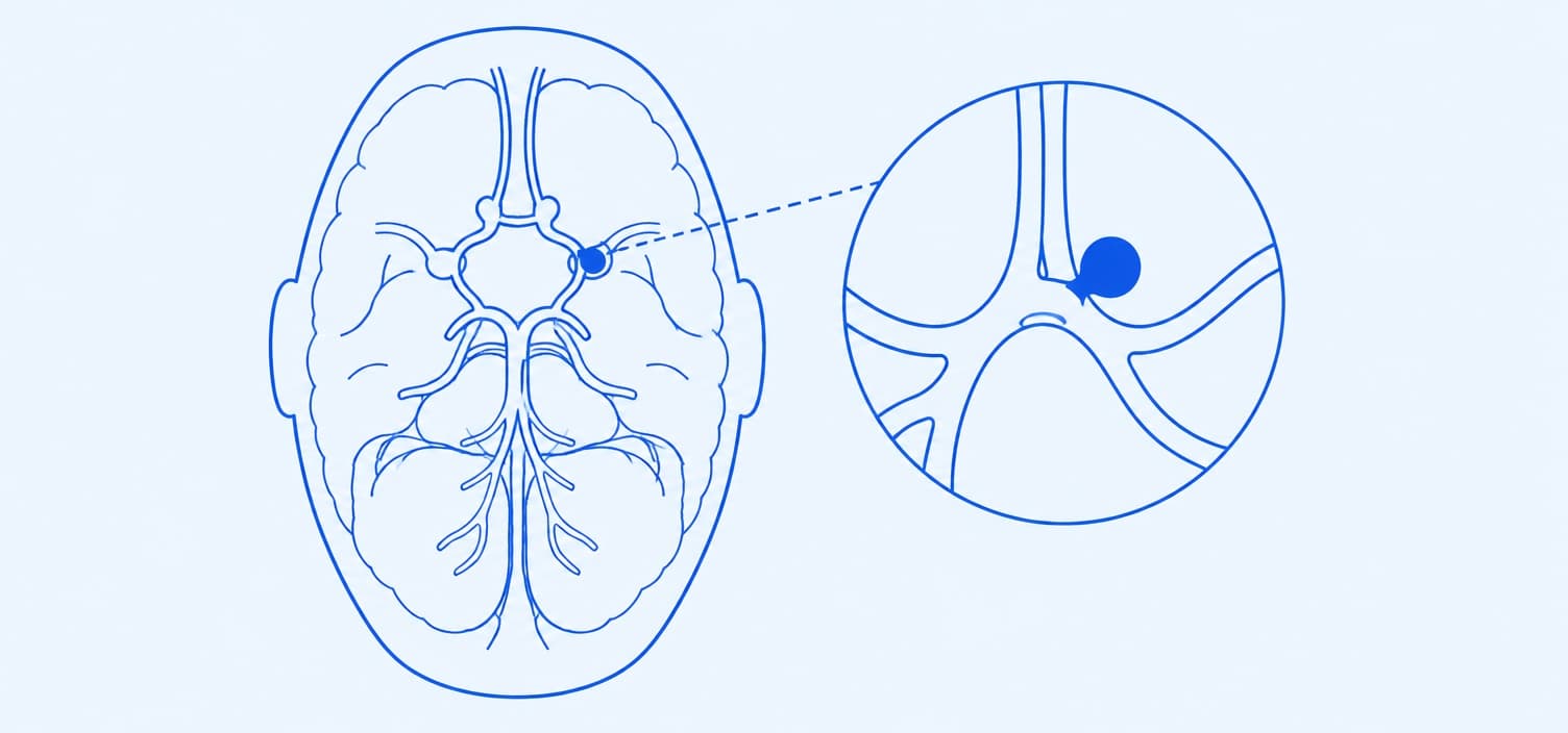
Brain aneurysm diagram showing a berry-shaped bulge on an artery at the base of the brain
A brain aneurysm: a weak spot in an artery wall at the base of the brain balloons out into a small sac that can leak or burst.

Most people assume an aneurysm forms overnight. It doesn't. Constant blood flow presses against a naturally thin point in an artery wall, and over years that spot stretches outward, the way a worn tyre bulges before it fails. Most brain aneurysms never rupture, and honestly, that surprises most people when they first hear it. The ones that do tend to sit where arteries branch at the base of the brain, where blood pushes hardest and turns most sharply.

Brain aneurysms become more common with age and show up more often in women than men. An AVM, by contrast, is usually present from birth. Not every brain aneurysm needs surgery. Many are simply watched, with regular scans tracking whether they change over time.

Types

Types of Brain Aneurysm

Your scan report will usually name one of the first three. The last card, an AVM, is a different blood vessel problem, but it's often confused with an aneurysm, so it helps to tell them apart.

Saccular Aneurysm

A round pouch bulging from one side of the artery, like a berry on a stem. Most aneurysms found on a scan look like this.

Fusiform Aneurysm

A bulge that widens the entire width of the artery rather than pouching to one side, often linked to high blood pressure or artery wall disease.

Mycotic Aneurysm

A rarer type caused by an infection weakening the artery wall from the inside, following a heart valve infection that reaches the brain's blood vessels.

AVM (Arteriovenous Malformation)

A tangle of arteries connected directly to veins, skipping the small vessels that slow blood down. It's usually present from birth, separate from an aneurysm.

Symptoms

Signs an Unruptured Aneurysm Can Cause

  • PERSISTENT HEADACHES

    A dull, one-sided headache that doesn't behave like your usual headaches and doesn't fully go away, sometimes the first sign a larger aneurysm is pressing on nearby tissue.

  • DROOPING EYELID

    One eyelid sagging lower than the other, caused by an aneurysm pressing on the nerve that controls the eye muscles, usually on just one side of the face.

  • DOUBLE OR BLURRED VISION

    Sudden trouble focusing or seeing two of something, especially when it comes on quickly rather than developing gradually the way normal eyesight changes do.

  • NUMBNESS ON ONE SIDE

    Tingling or weakness in the face on one side, a sign the aneurysm may be pressing against a nerve rather than just a blood vessel.

  • OFTEN NO SYMPTOMS

    Most unruptured brain aneurysms cause no symptoms at all and turn up only on a scan ordered for something else, which is why family history matters.

When To Worry

A Ruptured Brain Aneurysm Is Always An Emergency

A sudden, violent headache unlike anything you've felt before, often called the worst headache of your life, is the single most important warning sign. Don't wait to see if it passes. Call for emergency help immediately and get to a hospital with neurosurgery capability.

Call For Emergency Help If You Have:

Causes

Brain Aneurysm Causes

  1. 01

    High Blood Pressure

    Years of extra force against artery walls helps a weak spot stretch into an aneurysm, and it's the risk factor you can most easily control.

  2. 02

    Smoking

    Smoking damages the artery wall directly and is linked to both the formation of a brain aneurysm and a higher chance it eventually ruptures.

  3. 03

    Family History

    Having a parent or sibling with a brain aneurysm raises your own risk, and screening is usually discussed when two or more close relatives are affected.

  4. 04

    Connective Tissue Disorders

    Conditions like polycystic kidney disease or Ehlers-Danlos syndrome weaken artery walls throughout the body, making an aneurysm more likely to form, sometimes at a younger age.

Brain Aneurysm Risk Factors

  • AGE 30 TO 60

    Brain aneurysms are most often found in this age range, though they can form and rupture at any age.

  • SEX

    Women are diagnosed with a brain aneurysm roughly twice as often as men, particularly after menopause.

  • SMOKING & ALCOHOL

    Both raise the pressure and stress placed on artery walls, compounding the effect of any existing weak spot.

  • FAMILY HISTORY

    Having two or more affected parents, siblings or children is the clearest sign that screening scans are worth discussing.

Diagnosis

How Brain Aneurysm Is Diagnosed

  1. 01

    Initial Scan

    If you come in with a sudden severe headache or an aneurysm is suspected for another reason, a CT scan is usually the first test, since it's fast and widely available in an emergency.

  2. 02

    CT or MR Angiography

    A detailed scan that maps the brain's blood vessels using a contrast dye, showing the aneurysm's exact size, shape, and location well enough to plan treatment around it, without an invasive procedure.

  3. 03

    Cerebral Angiogram

    The gold standard test, a catheter threaded through a blood vessel captures the most detailed picture of an aneurysm, usually reserved for cases where surgery or coiling is already being planned.

Treatment

Brain Aneurysm & AVM Treatment Options

Treatment depends on the aneurysm's size, location, and whether it has ruptured, not on a single fixed rule. Many small, unruptured aneurysms are simply watched rather than treated right away.

  • WATCH AND SCAN

    is the approach for small, unruptured aneurysms with a low estimated risk of rupture, with imaging repeated on a schedule to track any change.

  • COILING

    is a catheter-based procedure that fills the aneurysm with tiny coils from inside the blood vessel, without opening the skull.

  • CLIPPING OR REMOVAL

    is open surgery to place a clip across the aneurysm's neck or remove an AVM directly, reserved for cases coiling can't safely reach.

Treatment Options Dr. Sunkara Offers

  1. 01

    Observation & Scanning

    NON-SURGICAL

    Regular MRI or CT angiography on a set schedule to track a small, unruptured aneurysm's size and shape over time, stepping in only if it changes.

  2. 02

    Endovascular Coiling

    NON-SURGICAL

    A catheter is guided up to the aneurysm, then filled with soft platinum coils that encourage clotting and seal it off from blood flow.

    See Neurovascular Surgery →
  3. 03

    Surgical Clipping

    SURGICAL

    A small metal clip is placed across the aneurysm's neck during open surgery, permanently closing it off while leaving the healthy artery intact.

    See Neurovascular Surgery →
  4. 04

    Stereotactic Radiosurgery for AVM

    NON-SURGICAL

    Targeted radiation delivered over one or a few sessions gradually closes off a small AVM's abnormal vessels, an option when open removal carries too much risk.

    See Stereotactic Radiosurgery →
Outlook

Is a Brain Aneurysm Curable?

Often, yes. Once coiling or clipping seals an aneurysm off, it rarely causes trouble again, though scans keep an eye on it over the years. A small one that's only being watched isn't cured, but many never grow or bleed. Many patients arrive expecting a ticking time bomb and leave with a calmer plan.

  • ABOUT 3 IN 100

    adults carry an unruptured brain aneurysm, and most never know because it never bleeds or causes a single symptom in their lifetime.

  • AFTER TREATMENT

    most coiled or clipped aneurysms stay sealed, though coiled ones need re-treatment a little more often, so follow-up scans matter.

  • IF IGNORED

    a sudden, severe headache waved off as a migraine delays care, and after a rupture, every hour before treatment counts.

Living With It

Living With a Treated or Watched Aneurysm

What can you actually control once you've been diagnosed with a brain aneurysm?

Dr. Raveesh Sunkara at his consultation desk, talking a patient through their condition
Before Your Appointment

Questions To Ask Your Doctor

  1. 01

    Is my aneurysm's size and shape considered high risk for rupture?

  2. 02

    Would you recommend watching this aneurysm, or treating it now?

  3. 03

    If treatment is needed, would coiling or clipping suit my case better?

  4. 04

    How often will I need follow-up scans if we choose to watch it?

  5. 05

    Should any of my close family members be screened as well?

Not Sure Which Option Is Right For You?

Bring your scan images to a consultation, and Dr. Sunkara will walk through what they mean for your specific case.

TESTIMONIALS

Trusted By Families Across India And Abroad.

★★★★★

Review summary

Her father had a grade 4 brain haemorrhage. Dr. Sunkara explained how critical it was, took every necessary step including a shunt, then guided his rehab.

Rushita G.

Brain haemorrhage

★★★★★

Review summary

Their parent was treated in an emergency and recovered after successful brain surgery. Dr. Sunkara even visited during rehab to check on progress.

Venkata Rama Reddy G.

Emergency brain surgery

★★★★★

Review summary

Her relative was admitted with a severe brain injury. Close monitoring and timely surgery led to a remarkable recovery, and he now leads a normal life.

Siri N.

Severe brain injury

FAQ

Your Brain Aneurysm Questions, Answered

A brain aneurysm is a weak, bulging spot on a blood vessel wall in the brain. Most stay silent for years and are found by accident, though a ruptured one is an emergency.

Get In Touch

Just Found Out You Have a Brain Aneurysm?

Bring your scan report for a second opinion, or book a consultation to talk through whether watching, coiling, or clipping is right for you.

Dr. Raveesh Sunkara

Written & reviewed by Dr. Raveesh Sunkara, Senior Consultant Neurosurgeon & Spine Surgeon · Last updated September 2026 · Next review due March 2027